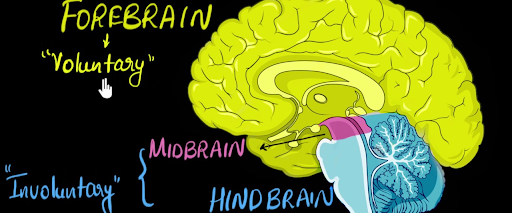
The division of the brain into forebrain, midbrain, and hindbrain represents a functional and developmental categorization that highlights distinct embryonic origins and broad functional roles. This division provides a broader organizational framework compared to other divisions that may focus on specific anatomical structures, neural circuits, or functional systems within the brain.
- Forebrain
- Midbrain
- Hindbrain
The forebrain is the largest and most complex division of the brain, encompassing the cerebral cortex, thalamus, hypothalamus, and limbic system. It plays a central role in various high-order cognitive functions, including thinking, reasoning, and perception. The cerebral cortex, forming the outer layer, is responsible for sensory and motor processing, as well as higher cognitive processes like language and problem-solving. The thalamus acts as a sensory relay station, while the hypothalamus regulates vital functions such as hunger, thirst, and body temperature. The limbic system, within the forebrain, is integral to emotional responses and memory formation. Dysfunction in the forebrain can impact a range of complex cognitive and emotional processes.
The midbrain, situated between the forebrain and hindbrain, serves as a critical relay station for sensory and motor pathways. It consists of the tectum, which includes the superior and inferior colliculi involved in visual and auditory reflexes, and the tegmentum, housing nuclei contributing to motor functions. The midbrain also contains the substantia nigra, playing a key role in motor control and implicated in conditions like Parkinson's disease. Additionally, the midbrain is crucial for arousal, attention, and the integration of sensory information, making it a vital component of the central nervous system.
The hindbrain, located at the posterior part of the brain, consists of critical structures such as the medulla oblongata, pons, and cerebellum. The medulla oblongata serves as the vital center for autonomic functions, including heart rate, respiratory rhythm, and reflex activities such as swallowing and vomiting. The pons acts as a bridge connecting different parts of the brain and is involved in sleep regulation and facial movements. The cerebellum, positioned beneath the cerebral hemispheres, plays a central role in coordinating voluntary muscle movements, maintaining balance, and fine-tuning motor skills. Dysfunction in the hindbrain can result in severe impairments in basic physiological functions and motor coordination.
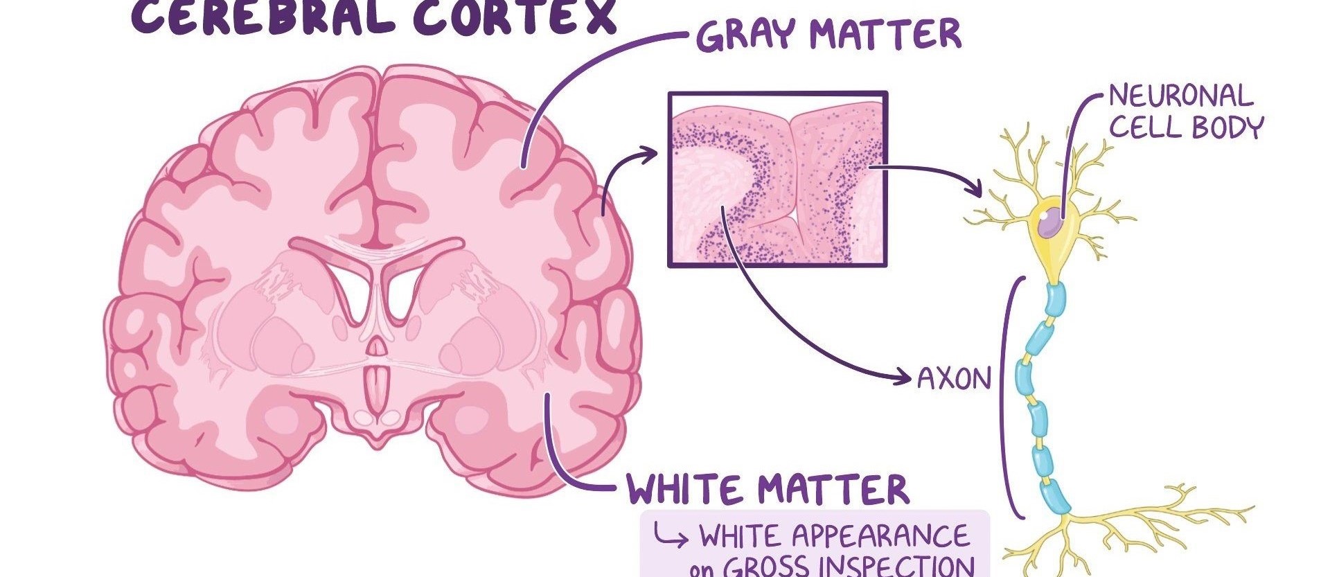
Gray and white matter are two different regions of the central nervous system. In the brain, gray matter refers to the darker, outer portion, while white matter describes the lighter, inner section underneath. In the spinal cord, this order is reversed: The white matter is on the outside, and the gray matter sits within.
- White Matter
- Grey Matter
White matter is a vital part of the central nervous system composed of myelinated nerve fibers, or axons, which facilitate communication between different regions of the brain and spinal cord. The myelin sheath, a fatty substance surrounding the axons, gives white matter its characteristic appearance. These fiber tracts form complex networks that enable the transmission of electrical signals, allowing for efficient and coordinated neural communication. White matter plays a crucial role in integrating information, coordinating motor functions, and facilitating the rapid transmission of signals across various brain regions, contributing to overall cognitive and motor functions.
Grey matter, a crucial component of the central nervous system, consists of neuronal cell bodies, dendrites, and glial cells. It forms the outer layer of the brain (cerebral cortex) and clusters in deeper regions, such as the brainstem and spinal cord. Grey matter plays a pivotal role in information processing, sensory perception, and motor function. Its high concentration of cell bodies contributes to the distinctive grey appearance and reflects its involvement in higher cognitive functions, including memory, emotions, and decision-making.
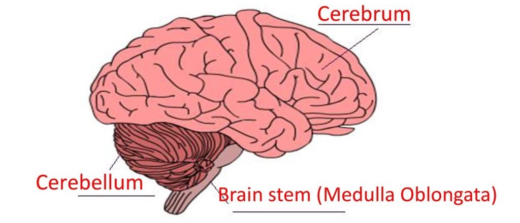
The division of the brain into the cerebrum, cerebellum, and brain stem represents a macroscopic distinction based on functional and anatomical characteristics. This division provides a broad organizational framework, distinct from other divisions that focus on microscopic structures, functional systems, or specific regions with specialized functions within the brain.
- Cerebellum
- Cerebrum
- Brain Stem
The cerebellum, located at the back of the brain, plays a crucial role in coordinating voluntary muscle movements and maintaining balance and posture. Comprising highly folded neural tissue, the cerebellum receives input from sensory systems and the cerebral cortex to fine-tune motor commands initiated by the cerebrum. Its intricate neural circuits facilitate precise and coordinated movements, ensuring fluidity in actions like walking, reaching, and speaking. Dysfunction of the cerebellum can lead to motor coordination deficits, impacting activities requiring precision and timing.
The cerebrum is the largest and most prominent part of the brain, constituting about 80% of its mass. It is divided into two hemispheres and is responsible for a multitude of higher cognitive functions, including sensory perception, motor control, language processing, and problem-solving. The outer layer, the cerebral cortex, is highly convoluted, increasing its surface area and accommodating intricate neural networks. The cerebrum plays a central role in complex mental processes, personality expression, and the integration of sensory and motor information.
The brain stem, situated at the base of the brain, is a vital region composed of three segments: the medulla oblongata, pons, and midbrain. It serves as a critical relay center, connecting the brain to the spinal cord and facilitating the passage of neural signals. The medulla controls involuntary functions such as breathing and heartbeat, while the pons aids in respiratory regulation and the transmission of signals between different brain regions. The midbrain plays a role in sensory processing, eye movement, and the coordination of motor activities. Together, the brain stem orchestrates fundamental life-sustaining functions and acts as a bridge between the brain and the rest of the body.
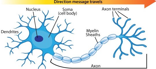
Neurons, the basic building blocks of the nervous system, consist of a cell body (soma), dendrites, and an axon. Dendrites receive signals from other neurons or sensory receptors, transmitting them to the cell body, which, in turn, processes the information and sends it down the axon for further communication with other neurons or effector cells.
- Dendrites
- Soma (Cell Body)
- Axon
Dendrites are branched extensions of a neuron that receive incoming signals from other neurons or sensory receptors. These structures play a critical role in information processing within the nervous system. The dendritic branches increase the surface area available for receiving signals, and the synaptic connections formed on dendrites allow for the transmission of neurotransmitters, enabling communication between neurons. The specific morphology and branching patterns of dendrites contribute to the unique characteristics of each neuron, influencing its ability to integrate and respond to incoming signals.
The soma, or cell body, is a crucial component of a neuron, containing the nucleus and other organelles essential for cellular functions. It integrates and processes incoming signals received from dendrites, determining whether the neuron will generate an action potential. The soma's role in information integration and cellular maintenance is fundamental to the overall function of the neuron in transmitting signals within the nervous system.
Axons are the elongated, thread-like extensions of neurons responsible for transmitting electrical signals away from the cell body. Encased in a myelin sheath formed by glial cells, axons facilitate the rapid conduction of nerve impulses. The axon's terminal branches communicate with other neurons or effector cells through synapses, allowing for the transfer of information across neural networks. Axons vary widely in length, with some extending over considerable distances within the nervous system.
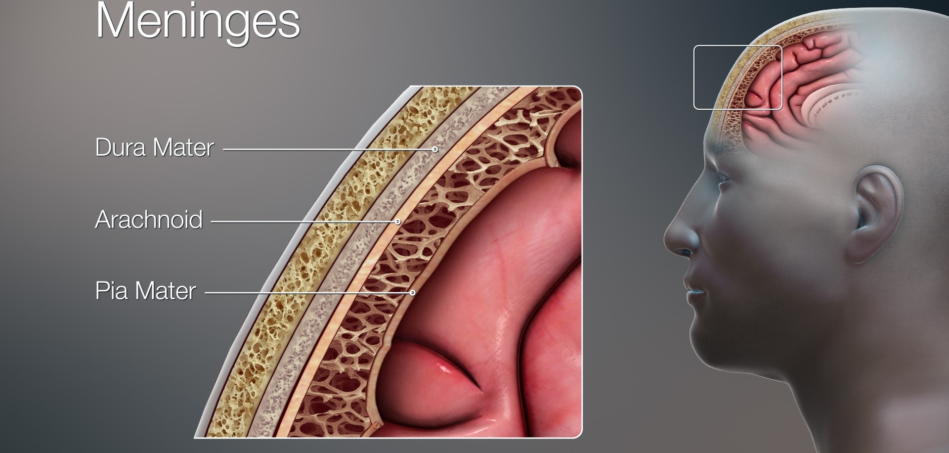
Meninges are the protective membranes surrounding the brain and spinal cord. Consisting of three layers—dura mater, arachnoid mater, and pia mater—meninges provide physical support, contain cerebrospinal fluid, and contribute to safeguarding the central nervous system from external trauma or infections.
- Dura
- Arachnoid
- Pia
The dura mater, the outermost layer of the meninges, is a durable and fibrous membrane that envelops the brain and spinal cord. It serves as a protective barrier, providing structural support and helping to maintain the shape of the brain. The dura mater contains blood vessels that supply oxygen and nutrients to the brain. Additionally, it forms the venous sinuses, channels that collect deoxygenated blood from the brain and facilitate its drainage back to the circulatory system.
The arachnoid mater is one of the three layers of the meninges, situated between the dura mater and the pia mater, enveloping the brain and spinal cord. This membrane, characterized by a spider-web-like appearance, forms a protective barrier and participates in cerebrospinal fluid circulation. Arachnoid granulations protrude into the dural venous sinuses, allowing for the drainage of cerebrospinal fluid back into the bloodstream. The arachnoid contributes to maintaining the delicate homeostasis of the central nervous system and acts as a barrier against potential pathogens or harmful substances.
The pia mater is the innermost layer of the meninges, directly adhering to the surface of the brain and spinal cord. Composed of delicate connective tissue, it follows the contours of the brain, entering its sulci and fissures. The pia mater is crucial for providing vascular support, as it carries blood vessels that nourish the underlying neural tissue. Additionally, it forms the outermost boundary of the subarachnoid space, where cerebrospinal fluid circulates, contributing to the overall protection and maintenance of the central nervous system.
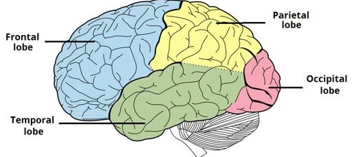
The brain hemisphere (parts of the cerebrum) has four sections, called lobes: frontal, parietal, temporal and occipital. Each lobe performs distinct cognitive functions, and their intricate interplay enables the brain to process a wide range of sensory and motor information, contributing to complex cognitive processes and behaviors.
- Frontal lobe
- Parietal lobe
- Occipital lobe
- Temporal lobe
The frontal lobe, positioned at the front of the brain, is pivotal for numerous higher cognitive functions and executive control. It houses the prefrontal cortex, responsible for decision-making, planning, and personality expression. The motor cortex in the frontal lobe coordinates voluntary muscle movements, while the Broca's area, typically located in the left hemisphere, is crucial for language production. Additionally, the frontal lobe plays a key role in emotional regulation, social behavior, and overall cognitive flexibility. Dysfunction in this area can impact personality, impulse control, and decision-making processes.
The parietal lobe, located near the top and back of the brain, is integral to various sensory and motor functions. It plays a key role in processing somatosensory information, enabling the perception of touch, pressure, temperature, and pain. Additionally, the parietal lobe is crucial for spatial orientation, contributing to an individual's awareness of their body's position in space. Higher cognitive functions, such as attention, calculation, and manipulation of objects in the environment, are also associated with the parietal lobe's intricate neural networks. Dysfunction in this area can lead to difficulties in spatial awareness, coordination, and sensory integration.
The occipital lobe, located at the back of the brain, is primarily responsible for visual processing and interpretation. It houses the primary visual cortex, where sensory input from the eyes is received and transformed into meaningful visual information. Additionally, the occipital lobe is involved in complex visual functions, such as object recognition, color perception, and the integration of visual stimuli. Lesions or damage to this lobe can lead to visual disturbances and impairments, emphasizing its pivotal role in the brain's overall sensory processing.
The temporal lobe, situated on each side of the brain above the ears, is primarily associated with auditory processing and memory functions. Within the temporal lobe lies the hippocampus, a crucial structure for the formation of new memories. Additionally, the temporal lobe plays a role in language comprehension and is interconnected with other brain regions involved in emotional regulation and the recognition of faces and objects. Dysfunction in the temporal lobe can lead to memory impairments, language difficulties, and challenges in processing auditory information.